

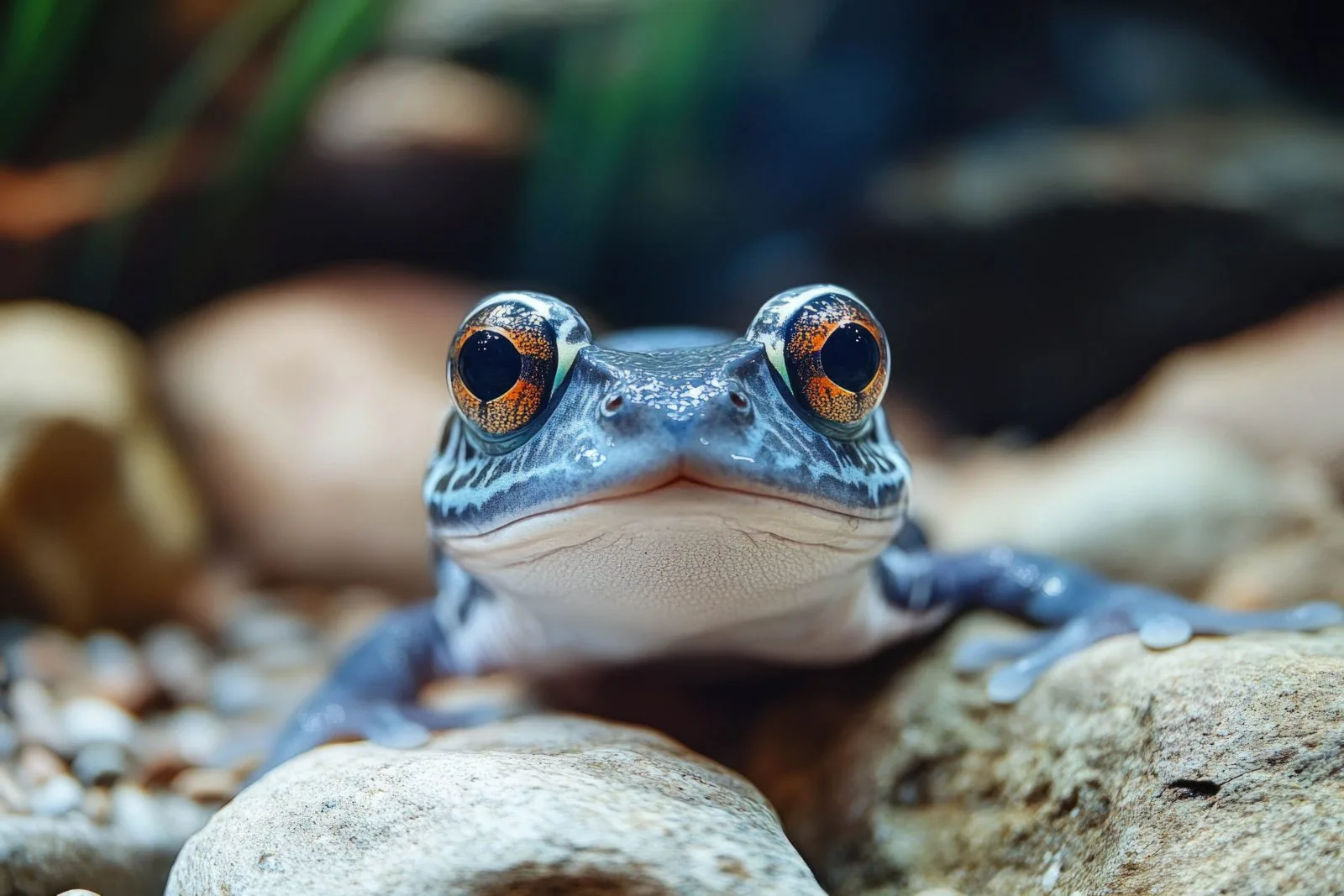
The research provides insights into biological mechanisms that could open new pathways for treating injuries in humans.
The ability to regenerate damaged tissues is one of the great hopes of modern medicine. In humans, this ability is very limited, and spinal cord injuries often cause permanent disabilities. However, some animals have developed surprising mechanisms that allow them to repair organs and body parts quickly and effectively, sparking intense scientific interest worldwide. One of these is the African frog, Xenopus laevis, which could be key to unraveling the mysteries of this process, opening doors to future therapies for spinal cord injuries and degenerative diseases.
Approximately a decade ago, Paula Slater, a native of Rancagua and now a researcher at the Department of Biological and Chemical Sciences of the Faculty of Science at Universidad San Sebastian, and an associate researcher at the Basal Center Ciencia & Vida, of the Fundacion Ciencia & Vida-Universidad San Sebastian, began working with this amphibian, a species that exhibits remarkable regenerative capacity in its early life stages. “It has many characteristics that make it an ideal model for studying both developmental biology and regeneration,” she notes. “As tadpoles, if the spinal cord is damaged or even if the tail is cut, it grows back and regains its function. But once they complete metamorphosis, they lose this ability,” Slater explains. This transition offers a unique opportunity to understand which biological mechanisms are activated during a successful regeneration event and which are deactivated during development, leading to a loss of regenerative capacity—a phenomenon that has barely been studied in humans.
In recent years, the team has made significant progress in understanding how the ‘powerhouses’ of cells, called mitochondria, react and change when damage occurs, and how these responses influence the recovery of the affected tissue.

Xenopus laevis in Chile
The African frog is not only a valuable model for research but is also an invasive species in Chile. Accidentally introduced in the 70s, Xenopus laevis established itself in the local environment and has affected native fauna. Although its presence in the local ecosystem generates negative impacts, its use in the laboratory remains fundamental for advancing scientific studies.
Interest in Xenopus laevis also lies in the fact that its development occurs outside the mother's body, facilitating direct observation and detailed study of its cellular and molecular processes in real-time. “In the case of frogs, it is external fertilization; therefore, the female lays the eggs in the water, and they are fertilized there. So everything happens externally, and that also allows us to study and work in early stages,” Slater details. This characteristic, uncommon in vertebrates, allows for precise analysis of how cells respond and contribute during development and the regeneration of nervous tissue.
One of the central lines of research at the neuroregeneration and metabolism laboratory, directed by Dr. Slater under the auspices of Centro Ciencia & Vida and Universidad San Sebastian, is understanding the role of mitochondria in regeneration. The researcher explains that “mitochondria regulate almost all cellular processes important for regeneration, such as cell division, proliferation, and differentiation. We have observed that in animals that regenerate, like this frog, mitochondria respond quickly to damage, altering their shape and function to protect cells and promote recovery.” Understanding these energetic and metabolic processes is fundamental to identifying which signals activate regeneration and how they could be replicated or stimulated in humans.
In her laboratory, they also investigate how the cellular environment, including the stiffness and electrical properties of the substrate where cells are located, influences regenerative capacity. Dr. Slater explains that “cells are not floating; they are on a substrate. This substrate has electrical components, stiffness, and specific molecules, which are altered after damage and also change during development.” Therefore, they are testing different substrates to better understand this interaction.
Regarding the time regeneration takes, Slater indicates that in tadpoles, specific damage to the spinal cord takes about 20 days to heal, recovering lost mobility. “In Xenopus, all regeneration is fast; as early as 6 hours, we see this adaptive response of the mitochondria to respond to the damage, and it begins to resolve by 24 hours,” she explains. Compared to slow human healing, this process is highly efficient, highlighting the importance of studying these models.
International Collaboration
The Chilean team works in close collaboration with international groups using different animal models to understand regeneration. She mentions the spiny mouse, a small African mammal with a remarkable, though slower, regenerative capacity. These comparative studies allow for the identification of which mechanisms are universal and which might be specific to certain species—information essential for developing therapies in humans. “We are collaborating in England, Portugal, and Germany with other groups working with different regeneration models,” Dr. Slater notes.
Although human regeneration is still far off, the accumulated knowledge opens the possibility of developing treatments that stimulate or reproduce these biological processes. Furthermore, advances in parallel fields such as electrical stimulation and nanotechnology are accelerating the search for therapies capable of regenerating damaged tissues. The associate researcher at Centro Ciencia & Vida and professor at Universidad San Sebastian explains that her goal is to identify factors that influence regeneration, such as the appropriate firmness of tissues or certain compounds that can have an impact.
The group's goal is to discover components and conditions that allow for the induction of regeneration in tissues that do not normally possess it, moving toward effective treatments for spinal cord injuries, multiple sclerosis, and other neurodegenerative pathologies. However, Dr. Slater warns that the transition from animal models to human clinical trials is long and complex. “Years of research, testing, and validation are needed, but every discovery brings us a little closer to that goal,” she acknowledges.
By: Richard García, Sandoval & Meirovich.