

An artificial intelligence analysis of more than 10 million cells enabled the identification of nine recurring patterns within different types of tumors.
An artificial intelligence analysis of more than 10 million cells allowed the identification of nine repeating patterns within different types of tumors.
These patterns, identified through massive observation of data from tissue biopsies, proved to be detectable in a simple blood sample, which would open a non-invasive pathway to monitor cancer.
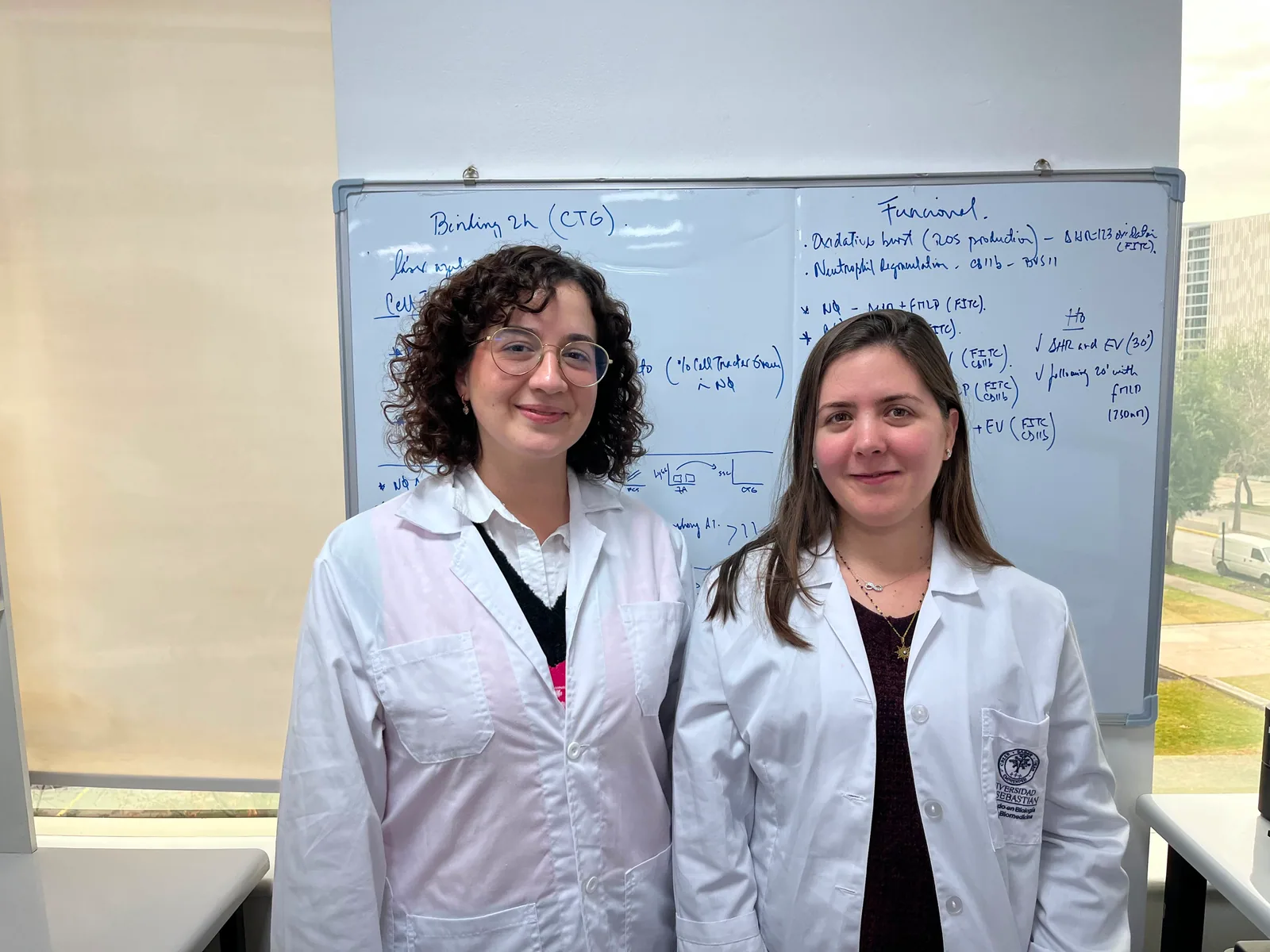
Two postdoctoral researchers from the Centro Basal Ciencia & Vida (Fundación Ciencia & Vida - Universidad San Sebastián) analyze the scope of this finding.
An artificial intelligence system identified nine spatial organization patterns (termed ecotypes) that repeat across different types of human tumors, based on the analysis of more than 10 million cells from over a hundred cases. The finding, published in the journal Nature, shows that these patterns can be detected in a blood sample instead of resorting to a surgical biopsy.
The study was developed by teams from Stanford University and the Chan Zuckerberg Biohub, with the participation of groups from Yale University and Washington University in St. Louis, and was published in 2026. In Chile, the research is analyzed by Dr. María José Oviedo and Dr. Laura Hernández, researchers at the tumor microenvironment and metastasis laboratory of the Centro Basal Ciencia & Vida (directed by Dr. Hugo González).
A tumor is not a homogeneous mass of cancer cells, state the postdoctoral researchers, whose work is framed within similar lines of research on the tumor microenvironment. Within it, immune cells, supporting cells, and blood vessels coexist, organized in spatial patterns that influence how the disease progresses and whether a specific treatment will be effective. Understanding this internal architecture is equivalent to understanding an entire ecosystem and not just its inhabitants.
“The tumor must be understood as a large city where not only tumor cells coexist, but also other cells that converse with the tumor and provide support for it to establish itself. This entire environment is known as the tumor microenvironment,” explains Dr. Hernández.
An artificial intelligence system identified nine spatial organization patterns (termed ecotypes) that repeat across different types of human tumors, based on the analysis of more than 10 million spatial transcriptomes and single-cell analyses from human carcinomas and melanomas. The finding, published in the journal Nature, shows that these patterns can be detected in a blood sample instead of resorting to a surgical biopsy.
Analyzing the tumor microenvironment requires dissociating the biopsy tissue, a process that yields the number and type of cells present but does not reveal how they relate to each other, nor where they are located within the tumor. Furthermore, a biopsy captures only a limited and specific fragment, insufficient to represent the heterogeneity of an entire tumor mass. Spatial transcriptomics is the technique that allows solving this puzzle and studying the neighborhoods established within the tumor microenvironment, keeping the cells exactly in their original position within the tissue.
Nine patterns, a common map
The study started with a simple question: What makes a tumor respond or not to a treatment? To answer it, the teams from Stanford University and the Chan Zuckerberg Biohub gathered spatial transcriptomics data from more than 10 million individual cells, originating from over a hundred human tumors—such as carcinomas and melanomas—and trained a machine learning system to recognize repeating organizational patterns within these tumors.
Dr. Oviedo, who at the Centro Basal Ciencia & Vida works mainly with animal models and human tissue samples, closely followed this line of research. “Artificial intelligence is combined with single-cell spatial transcriptomics data from more than 10 million samples to identify the spatial arrangement of these cells in the tumor,” the researcher details.
The analysis was not limited to counting how many cells of each type were in the tumor. It sought to determine where they were located relative to each other—tumor cells, immune cells, blood vessels, and supporting tissue—and how this spatial arrangement related to the clinical outcome of each patient.
The result was nine spatial ecotypes that consistently repeat across different types of carcinomas and melanomas, each with a characteristic biology and location within the tumor. Several of these were clearly associated with response to immunotherapy and patient survival. “They discovered nine ecotypes that correlated with DNA methylation profiles; in the laboratory, working at the bench, it would be very difficult to detect these patterns, but through these tools, it becomes much easier,” adds Dr. Oviedo.
From biopsy to blood
Until now, access to this information was very limited, as obtaining biopsies is an invasive procedure that provides a partial and snapshot-like picture of tumor development. The techniques used today require tissue dissociation to determine the number and type of cells, but not how they relate to each other, nor where they are located.
Dr. Hernández, who at the Centro leads a line of work with samples from breast cancer patients, within the framework of a collaboration with the Fundación Arturo López Pérez (FALP), explains the central contribution of the study. “The highlight of this paper is that this common pattern, these identified genomic marks, could be detected in clinical practice using a blood sample. Furthermore, it could predict whether the patient might respond well to therapy or if they have a shield that will block the treatment.”
The finding consisted of proving that the nine ecotypes identified in tumor tissue—previously invisible—are also recorded in DNA fragments that tumors release into the bloodstream, known as circulating cell-free DNA. This possibility changes the logic of sample representativeness.
A conventional biopsy captures a limited fragment of the tumor at a specific moment; a blood sample, on the other hand, can more comprehensively reflect the entirety of a patient's tumor lesions, including those that were not directly biopsied. “A blood sample can capture information from different tumor lesions, whereas a biopsy is taken at a specific time and is very small, not representing the entire tumor,” indicates Dr. Oviedo.
A compass for immunotherapy
In a group of about 100 patients with advanced melanoma, the levels of these ecotypes measured in blood before starting immunotherapy were robustly associated with treatment response and survival, according to the study published in Nature.
For Dr. Laura Hernández, this correlation has a direct practical consequence if it can be confirmed in larger studies. “If this can be validated in different types of tumors, it could prevent subjecting patients to therapies to which they would not respond, and which generate multiple adverse effects. Additionally, it would allow dynamic monitoring, through the blood sample, of when the tumor might be changing its ecotype from sensitive to resistant or vice versa, in order to adapt treatment approaches,” the researcher suggests.
Immunotherapy and other oncological treatments are not without adverse effects. Identifying beforehand which patients are less likely to respond would allow redirecting the therapeutic strategy before exposing them to a treatment that will not benefit their condition. The study's authors themselves, however, put the finding into perspective: it is basic research, not a test available in clinics or hospitals.
Before becoming a diagnostic tool, the results must be validated in larger cohorts and in different types of cancer, the specialists agree. At the Centro Basal Ciencia & Vida, both researchers continue to explore the relationship between the spatial arrangement of tumor cells, both in animal models and in patient samples.
Text by Luis Francisco Sandoval, Agencia S&M Comunicaciones.